

Principle of Fluorescence Microscopy
- A fluorescence microscope uses fluorescence to generate an image.
- The specimen would be first prepared by injecting the fluorophores, which are fluorescence indicators such as Green fluorescent protein (GFP) or Oregon Green™ 488 BAPTA-1, AM (OG488).
- After that, it would be illuminated with specific wavelengths of light which is absorbed by the fluorophores.
- When the fluorophores absorb a high-energy photon (from the light source), the chemical system would be excited electronically and vibrationally, and become unstable at high energy levels.
- This unstable chemical system would soon release the absorbed photon energy through non-radiative transition first and eventually emitted fluorescence at a longer wavelength.
- The emitted fluorescence would be separated from the illumination light source through the emission filter, and the camera would capture the image from the specimen.
- Through the fluorescence microscope, we could conduct an observation of the specific structures or activities which have been labeled for fluorescence only.
 |
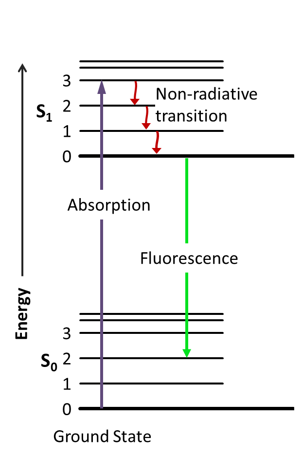 |
| FIG. 1. The working principle of Fluorescence Microscopy. The arc lamp together with the blue excitation act as the incident light source with specific wavelengths. GFP is used as the fluorophores. | FIG. 2. Jablonski diagram including the energy level of the fluorophore. The photochemical reaction includes the absorption, non-radiative transition, and emission of fluorescence. |